



KEYWORDS: HIV, Advanced HIV disease, Neurosyphilis, Disseminated cryptococcosis, Herpes genitalis, Polymicrobial infections
INTRODUCTION
People living with HIV (PLHIV), particularly those with advanced HIV disease, remain highly vulnerable to opportunistic infections (OIs) as a consequence of profound and prolonged immune dysregulation1. Late diagnosis, poor linkage to care, treatment interruption, and late presentation continue to be associated with advanced disease states despite the widespread availability of antiretroviral therapy, especially in resource-limited settings. In these patients, OIs are often the earliest clinical manifestation of HIV infection or advanced immunodeficiency and remain a major cause of hospitalization and death. Importantly, OIs in advanced HIV often present atypically, disseminated or severely, reflecting pathogen burden and impaired host immune response1.
The occurrence of polymicrobial infections in PLHIV introduces an additional layer of complexity to both diagnosis and management. Concurrent infections may present with overlapping or nonspecific clinical features, obscure classical disease patterns, and evolve asynchronously, leading to missed or delayed diagnoses. Therapeutic decision-making is further complicated by drug–drug interactions, cumulative toxicities, immune reconstitution inflammatory syndrome, and competing priorities in sequencing antimicrobial therapy 2, 3. While individual opportunistic infections such as cryptococcosis, syphilis and Herpes simplex virus infection are well recognized in advanced HIV, their simultaneous occurrence is rare and under-reported. Furthermore, literature has often focused on single-pathogen paradigms, leaving large gaps in our knowledge of the clinical behaviour, diagnostic pitfalls, and outcomes of true polymicrobial opportunistic disease in severely immunocompromised hosts4.
We describe a rare and diagnostically challenging case of concurrent disseminated cryptococcosis, neurosyphilis and herpes simplex virus infection in a patient with advanced HIV. This case illustrates clinical and therapeutic dilemmas in the simultaneous management of multiple OIs, and raises interesting questions about the biology and interaction of co-infecting pathogens within the same host.
The possible interactions of microbes, either synergistic or antagonistic, symbiotic or parasitic, mutualistic or commensal are poorly understood in the context of severe immunodeficiency and clinical practice. This unusual constellation of infections is detailed in order to highlight gaps in current diagnostic approaches, describe need for heightened clinical suspicion of polymicrobial disease in advanced HIV, and contribute to the evolving understanding of pathogen–pathogen interactions in immunocompromised hosts.
CASE PRESENTATIONS
A 28-year-old male from North India, with a history of occasional smoking and alcohol consumption, presented with a painful genital ulcer and lip rash of approximately 2.5 months’ duration, accompanied by intermittent fever for 2 months and vomiting for 2 days. He had been recently diagnosed as HIV-reactive at an outside healthcare facility and had not yet initiated antiretroviral therapy.
On day 0, during the course of evaluation, the patient developed acute-onset right-sided hemiplegia, slurred speech, facial asymmetry, and altered sensorium. Initial neuroimaging performed at presentation was suggestive of an acute cerebrovascular event, and a provisional diagnosis of stroke was considered.
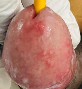
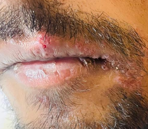
Cutaneous examination revealed multiple painful vesicular and erythematous lesions with superficial erosions over the glans penis (Fig. 1A), consistent with genital herpes, as well as grouped vesicular and crusted lesions over the upper and lower lips (Fig. 1B), characteristic of herpes labialis.
Baseline and follow-up investigations are summarized in Table 1. The patient had advanced immunosuppression with a CD4 count of 51 cells/µL. Serum RPR was positive at presentation and subsequently became non-reactive following antimicrobial therapy. Serum cryptococcal antigen remained repeatedly positive during the diagnostic course.
Table 1: Summary of investigations during hospitalisation and follow-up of the patient.
CSF analysis showed pleocytosis (20 cells, 100% monomorphs), elevated protein (355 mg/dl), and low glucose (45 mg/dl; corresponding RBS 185 mg/dl) but sterile cultures, negative Gram stain, negative Cryptococcal antigen, negative GeneXpert , negative acid fast staining, and no fungal elements on KOH mount, and negative Indian ink staining. CSF VDRL test was not performed because it was not available. Magnetic resonance imaging (MRI) brain revealed multiple ring-enhancing lesions initially diagnosed as tuberculomas but later reinterpreted as cryptococcomas after clinic-radiological correlation. Contrast-enhanced computed tomography (CECT) thorax revealed tree-in-bud opacities, but no microbiological or radiological evidence of active pulmonary tuberculosis was seen.
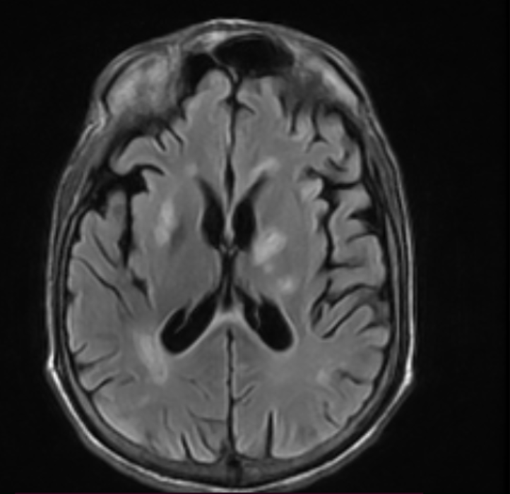
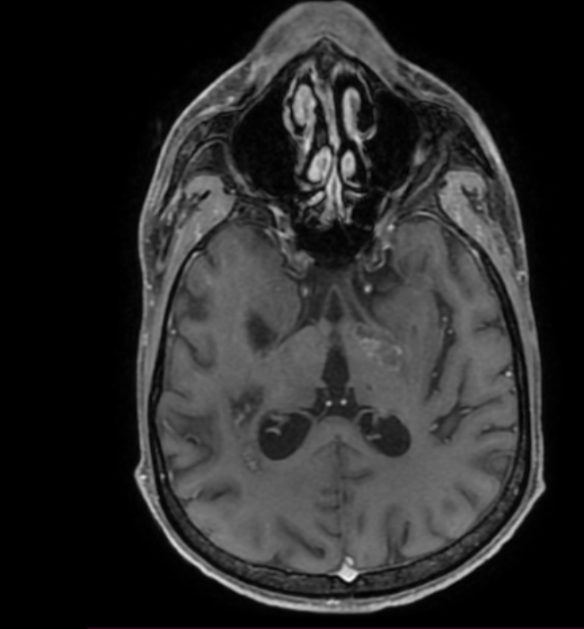
Further testing revealed positive serum cryptococcal antigen. MRI brain (Fig. 1C, T2 FLAIR (Pre-contrast) and 1D, Post-contrast T1) revealed multiple ring enhancing lesions involving bilateral gangliocapsular regions, left thalamus and right periventricular area. Initially, these lesions were diagnosed as tuberculomas. However, repeat neuroimaging, persistently positive serum cryptococcal antigen testing and multidisciplinary discussion led to reclassification of the lesions as cryptococcomas.
[A] [B]
[C] [D]
Figure 1: Clinical photographs & Contrast-enhanced MRI brain images. (A) showing multiple grouped vesicular and erythematous lesions with superficial erosions over the glans penis, consistent with genital herpes (herpes genitalis). Lesions are present at varying stages of healing. (B) showing grouped vesicular and crusted lesions involving the upper and lower lips, localized to the vermilion border, consistent with herpes labialis caused by reactivation of herpes simplex virus type 1 (HSV-1). Contrast-enhanced MRI brain showing multiple variable-sized, relatively well-defined, round to ovoid-shaped hyperintense lesions, seen in bilateral gangliocapsular, left thalamus, and right periventricular region (C, T2 FLAIR). A few of them are showing post-contrast peripheral ring enhancement on the T1 image (D).
Given the presence of a genital ulcer and a positive serum rapid plasma reagin (RPR) test, neurosyphilis was also considered in the differential diagnosis. CSF analysis supported central nervous system involvement, though cultures remained sterile.
The patient was managed with a multidisciplinary approach and received:
Liposomal amphotericin B and flucytosine as induction therapy for cryptococcosis, followed by fluconazole consolidation
Ceftriaxone and benzathine penicillin G (due to the lack of availability of aqueous penicillin) for neurosyphilis
Acyclovir for herpes labialis and genitalis
Broad-spectrum antibiotics for secondary bacterial infections
Trimethoprim-sulfamethoxazole led to drug-induced pancytopenia and was discontinued; any other drug could not be given as prophylaxis against PCP, as Atovaquone was unavailable, and Dapsone was not tried due to anaemia.
Antiretroviral therapy (ART) was initiated at an outside hospital facility where the patient was admitted before coming to our institute; however, on admission to our hospital, we discontinued the ART due to the presence of opportunistic infections, including cryptococcal meningitis, and planned to reintroduce it on follow-up after 6 weeks of antifungal therapy.
Supportive care, including nutritional support and physiotherapy
His hospital course was complicated by aspiration pneumonia, catheter-associated urinary tract infection, septic shock and drug induced pancytopenia. Despite these complications, the patient improved gradually neurologically and systemically with appropriate antimicrobial therapy and supportive care. However, there was residual right-sided hemiplegia at discharge.
The patient was discharged on fluconazole consolidation therapy, acyclovir and supportive management. He was advised to have close outpatient follow-up including repeat laboratory monitoring, neuroimaging and continued physiotherapy for neurological rehabilitation.
DISCUSSION
This case highlights the complex clinical, diagnostic, and therapeutic challenges encountered in advanced HIV disease (PLHIV) complicated by simultaneous polymicrobial opportunistic infections, namely disseminated cryptococcosis, neurosyphilis, and herpes simplex virus infection. The patient presented with profound immunosuppression (CD4 count 51 cells/µL) and overlapping systemic, neurological, and mucocutaneous manifestations, illustrating how multiple infections can coexist, evolve in parallel, and obscure classical disease patterns. Such presentations reinforce the need for a broad, syndromic, and iterative diagnostic approach rather than reliance on a single unifying diagnosis, particularly in late-presenting PLHIV.
Managing PLHIV with multiple opportunistic infections remains a major clinical challenge, especially in resource-limited and tuberculosis-endemic settings. Rapid screening for OIs, prophylaxis, prompt initiation of antiretroviral therapy (ART), and intensified adherence support are major recommendations from the World Health Organization (WHO) guideline1. OIs such as cryptococcosis, tuberculosis, histoplasmosis, and herpesvirus infections contribute substantially to HIV-related morbidity and mortality. Evidence suggests that rapid , routine screening for these infections is feasible and results in improved clinical outcomes in hospitalised PLHIV patients with very low CD4 counts5. Also, timely initiation of primary and secondary prophylaxis is critical for preventing both initial and recurrent OIs, with discontinuation only when sustained immune recovery on ART occurs6.
The coexistence of neurosyphilis, disseminated cryptococcosis, and herpes simplex infection in this patient reflects the depth of immune dysfunction associated with advanced HIV. While polymicrobial infections are not uncommon in PLHIV, the specific symbiosis observed in this patient is rare7. One of the most significant diagnostic challenges encountered was differentiating cryptococcomas from tuberculomas, a recurring dilemma in tuberculosis-endemic regions like ours. Both diseases present with multiple ring-enhancing lesions on neuroimaging and overlapping clinical features, including fever, focal neurological deficits, and seizures8, 9. Cryptococcal granulomas can closely mimic tuberculomas on MRI, with variable imaging appearances that depend on disease stage, as seen with tuberculous brain involvement. While a cryptococcoma almost universally appears hyperintense on T2-weighted images, a tuberculoma can show either hyperintensity or hypointensity on T2, depending on its stage of illness. Reports from South India have described cases initially diagnosed as tuberculosis on imaging but later confirmed as cryptococcosis on tissue examination10. In our patient, persistently positive serum cryptococcal antigen testing and lack of evidence supporting active tuberculosis favored a diagnosis of cryptococcosis.
Cryptococcus neoformans remains a leading cause of fungal meningitis globally and a major contributor to HIV-associated mortality, particularly in sub-Saharan Africa11,12. WHO estimates indicate that cryptococcal meningitis accounts for over 180,000 deaths annually among PLHIV13. Cryptococcomas represent a localized granulomatous form of disease and pose significant diagnostic challenges because they radiologically resemble tuberculomas9. In this case, longitudinal clinical assessment, repeat antigen testing, and multidisciplinary review were critical in guiding appropriate antifungal therapy.
Neurosyphilis, previously considered uncommon in the ART era, has re-emerged among PLHIV, particularly in those with delayed diagnosis or poor engagement in care14. Clinical manifestations are variable, from meningitis to meningovascular disease, and may present as stroke. In this patient, syphilis likely contributed to the neurological presentation alongside cryptococcosis. Early recognition and treatment with ceftriaxone and benzathine penicillin remain essential to prevent irreversible neurological damage15.
Herpes simplex virus (HSV) infection is also one of the most frequent OIs in PLHIV3. In advanced disease, HSV often presents with chronic, extensive, or atypical mucocutaneous lesions. Prompt initiation of acyclovir led to the resolution of lesions in our patient; however, HSV infection further exemplifies the symbiotic infectious burden borne by severely immunocompromised individuals.
Therapeutic management in such cases is further complicated by drug–drug interactions and cumulative toxicities. Agents used for cryptococcosis, neurosyphilis, HSV infection, bacterial infections, and ART all carry potential adverse effects. Our patient developed drug-induced pancytopenia, necessitating modification of therapy, with resolution following discontinuation of trimethoprim–sulfamethoxazole16. Supportive care measures, such as nutritional supplementation and physiotherapy, played a major role in recovery, alongside monitoring for adverse drug reactions.
The overlapping presentation and symbiosis of multiple OIs in this patient highlight the limitations of classical diagnostic heuristics and the concept of ‘Occam’s razor’, in which fewest possible set of explanations should be considered in presence of multiple explanations for same phenomenon. In these situations, the value of multidisciplinary collaboration between neurology, infectious diseases, microbiology, radiology and rehabilitation services is stressed to maximize patient outcomes17,18.
Similar reports of advanced HIV/PLHIV complicated with multiple OIs have been described in literature, often involving viral, bacterial, fungal and protozoal pathogens leading to high morbidity and mortality4. A case report published in 2017 described concurrent neurosyphilis, cryptococcal meningitis and tuberculous meningitis in an HIV-infected patient, highlighting the need to actively evaluate for multiple CNS infections even after establishing the first diagnosis19. In another case from the United States, concurrent cryptococcal meningitis and neurosyphilis were reported in an HIV patient7. These observations suggest that diagnostic anchoring should be avoided in advanced HIV. Beyond diagnostic overlap and therapeutic complexity, this case also raises important considerations regarding polymicrobial interactions within the immunocompromised host. In advanced HIV, severe immune dysregulation alters host–pathogen dynamics, permitting simultaneous persistence of multiple organisms that would otherwise be controlled or competitively excluded. Emerging evidence suggests that mechanisms of immune modulation, niche sharing, metabolic cooperation, or immune diversion, potentially influencing disease severity, tissue tropism, and response to therapy, are present where co-infecting pathogens interact through20, 21. For example, chronic viral infections such as herpes simplex virus may disrupt mucocutaneous barriers and local immunity, facilitating secondary infections, while systemic fungal or bacterial infections may further impair cell-mediated immune responses critical for pathogen clearance. But the exact nature of these interactions, be they synergistic, antagonistic or just permissive, is poorly defined in clinical settings. This case highlights the importance of moving away from single-pathogen frameworks and considering the polymicrobial ecology of the human host, especially in patients with advanced HIV disease.
Limitations of this report include the single patient design that limits generalizability. Clinical limitations did not allow the tissue diagnosis of intracranial lesions. Microbiological confirmation was based on serological and radiological correlation and not histopathology. Follow-up was also limited, preventing a complete evaluation of long-term neurologic and functional outcomes. This case, however, is an example of the diagnostic complexity, therapeutic challenges and the need for integrated care in patients with advanced HIV and concomitant polymicrobial opportunistic infections.
CONCLUSIONS
This case report illustrates the clinical and diagnostic challenges of advanced HIV disease complicated by concomitant polymicrobial opportunistic infections. Disseminated cryptococcosis, neurosyphilis and herpes simplex virus infection occurring simultaneously exemplify the degree of immunosuppression which permits co-infection with the potential for clinical, radiological and pathophysiological overlap beyond the scope of single disease diagnostic paradigms. This case highlights the need to take into account the dynamic interplay between co-infecting pathogens in the immunocompromised host, as well as the importance of high clinical suspicion, iterative diagnostic reassessment, and multidisciplinary management. Early recognition, prompt initiation of targeted antifungal, antimicrobial, and antiretroviral therapy, vigilant surveillance for treatment-related complications, and supportive care continue to be fundamental to improving outcomes and reducing morbidity in patients with advanced HIV.
ACKNOWLEDGEMENT
None
CONFLICTS OF INTEREST STATEMENT
None
SOURCE OF FUNDING
None
CONSENT AND ETHICAL APPROVAL
Informed consent was obtained, but there is an ethics waiver as per the institute protocol.
AUTHOR’S CONTRIBUTION
Both authors contributed equally to the data collection and manuscript writing.
AI DECLARATION
No AI tool was used to generate research data, perform data analysis, interpret findings, formulate scientific conclusions, or make editorial decisions. All AI-assisted content was critically reviewed, verified, and revised by the authors. The authors take full responsibility for the accuracy, originality, integrity, and final content of the manuscript.
REFERENCES
World Health Organization. The Advanced HIV Disease Research Landscape. Geneva: WHO; February 19, 2024. Accessed February 2026. https://www.who.int/publications/i/item/9789240089020
Ford N, Shubber Z, Meintjes G, et al. Causes of hospital admission among people living with HIV worldwide: a systematic review and meta-analysis. Lancet HIV. 2015;2(10):e438-e444. https://doi.org/10.1016/s2352-3018(15)00137-x
Panel on Opportunistic Infections in Adults and Adolescents with HIV. Guidelines for the Prevention and Treatment of Opportunistic Infections in Adults and Adolescents with HIV. Department of Health and Human Services; 2023. https://doi.org/10.1037/e545852006-001
Boozhmehrani MJ, Hoseinnejad A, Lesani S, et al. Comprehensive management of HIV with multiple opportunistic infections: a case report. Clin Case Rep. 2025;13(10):e71216. https://doi.org/10.1002/ccr3.71216
Pasqualotto AC, Sued O, Reis N, Silva LR, Soares RBA, Godoy CSM, et al. Impact of the introduction of a package of care involving early detection of opportunistic infections, a prospective multicenter cohort study of people living with HIV/AIDS in Brazil. Lancet Reg Health Am [Internet]. 2025;45(101085):101085. Available from: http://dx.doi.org/10.1016/j.lana.2025.101085
Pasqualotto AC, Sued O, Reis N, Silva LR, Soares RBA, Godoy CSM, et al. Impact of the introduction of a package of care involving early detection of opportunistic infections, a prospective multicenter cohort study of people living with HIV/AIDS in Brazil. Lancet Reg Health Am [Internet]. 2025;45(101085):101085. Available from: http://dx.doi.org/10.1016/j.lana.2025.101085
Swe T, Laurent B, Shah N. Concurrent central nervous system infective pathology in a severely immunocompromised patient. J Family Med Prim Care. 2016;5(3):685. https://doi.org/10.4103/2249-4863.197304
Berkowitz AL. Approach to neurologic infections. Continuum (Minneap Minn). 2021;27(4):818-835. https://doi.org/10.1212/con.0000000000000984
Shetty G, Avabratha KS, Rai BS. Ring-enhancing lesions in the brain: a diagnostic dilemma. Iran J Child Neurol. 2014;8(3):61-64. https://pmc.ncbi.nlm.nih.gov/articles/PMC4135283/
Patro SN, Kesavadas C, Thomas B, Kapilamoorthy TR, Gupta AK. Uncommon presentation of intracranial cryptococcal infection mimicking tuberculous infection in two immunocompetent patients. Singapore Med J. 2009;50(4):e133-e137. https://pubmed.ncbi.nlm.nih.gov/19421667/
Rajasingham R, Smith RM, Park BJ, et al. Global burden of disease of HIV-associated cryptococcal meningitis: an updated analysis. Lancet Infect Dis. 2017;17(8):873-881. https://doi.org/10.1016/s1473-3099(17)30243-8
Lawn SD, Bekker LG, Myer L, Orrell C, Wood R. Cryptococcal immune reconstitution disease: a major cause of early mortality in a South African antiretroviral programme. AIDS. 2005;19(17):2050-2052. https://doi.org/10.1097/01.aids.0000191232.16111.f9
World Health Organization. Guidelines for Diagnosing, Preventing and Managing Cryptococcal Disease Among Adults, Adolescents and Children Living with HIV. Geneva: WHO; 2022. https://pubmed.ncbi.nlm.nih.gov/35797432/
Chen HJ, Yu T, Peng J. Asymptomatic neurosyphilis may need early intervention in people living with HIV. Chin Med J (Engl). 2020;133(23):2877-2879. doi:10.1097/CM9.0000000000001143 https://doi.org/10.1097/cm9.0000000000001143
Hamill MM, Ghanem KG, Tuddenham S. State-of-the-art review: neurosyphilis. Clin Infect Dis. 2024;78(5):e57-e68. doi:10.1093/cid/ciad437 https://doi.org/10.1093/cid/ciad437
Parajuli P, Ibrahim AM, Siddiqui HH, Lara Garcia OE, Regmi MR. Trimethoprim-sulfamethoxazole induced pancytopenia: A common occurrence but A rare diagnosis. Cureus [Internet]. 2019; Available from: http://dx.doi.org/10.7759/cureus.5071
Shah N, Owen L, Bhagani S. “Occam’s scissors”: opportunistic infections in advanced HIV infection. BMJ Case Rep. 2013. https://doi.org/10.1136/bcr-2013-009544
Pinheiro MVC, Ho YL, Nicodemo AC, Duarte-Neto AN. The diagnosis of multiple opportunistic infections in advanced stage AIDS: when Ockham’s razor doesn’t cut it. Autops Case Rep. 2018;8(2):e2018028. doi:10.4322/acr.2018.028 https://doi.org/10.4322/acr.2018.028
Gonzales Zamora JA, Espinoza LA, Nwanyanwu RN. Neurosyphilis with concomitant cryptococcal and tuberculous meningitis in a patient with AIDS: report of a unique case. Case Rep Infect Dis. 2017;2017:4103858. doi:10.1155/2017/4103858 https://doi.org/10.1155/2017/4103858
Deol P, Miura TA. Respiratory viral coinfections: interactions, mechanisms and clinical implications. Nat Rev Microbiol. 2025;23(12):757-770. doi:10.1038/s41579-025-01225-3 https://doi.org/10.1038/s41579-025-01225-3
Das D, Panda PK. Polymicrobial infection in COPD exacerbation. JASPI. 2024;2(4 Suppl). https://doi.org/10.62541/jaspi122024